Wednesday, 14 November 2018

Dedifferentiation and Re differentiation

What is Dedifferentiation?

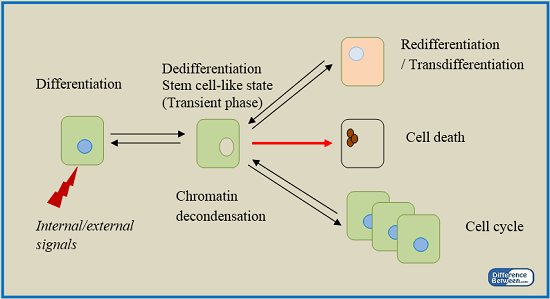
Under certain conditions, plants cells that are already differentiated and lost the ability of further division regain the capacity of division and differentiation. This process is known as dedifferentiation. The fully differentiated parenchyma cells undergo dedifferentiation, which leads to the formation of cork cambium and inter-fascicular cambium. A dedifferentiated tissue has the ability to act as meristem that could give rise to a different set of cells. The ability of those cells for further differentiation depends on different parameters such as genetic and epigenetic variations. This concept is used in plant tissue culture to develop a callus.

What is Redifferentiation?

Once new cells are formed from the dedifferentiated tissues that act as meristems, the cells lose their ability for further division and differentiation. Eventually, they get mature in order to accomplish specific functions of the plant body. Secondary xylem and secondary phloem are the best examples to describe the process of redifferentiation. Dedifferentiated vascular cambium divides further to give rise to the secondary xylem in the inside and secondary phloem on the outside. The secondary phloem and secondary xylem cells lose their ability for further division; instead, they become mature to fulfill specific functions of the plant body, which include transportation of food and water, respectively. Phelloderm is a layer of secondary tissues that is produced by the dedifferentiated cork cambium. Similar to secondary xylem and phloem, phelloderm’s cells lose their ability for further differentiation but become mature in order to perform specific functions such as limitation of dehydration and prevention of the entry of pathogens into the plant body due to the destruction of the epidermis.
Difference Between Dedifferentiation and Redifferentiation
Figure 01: Differentiation and Redifferentiation

Cytodifferentiation


Cytodifferentiation
The cells of some callus mass fre­quently differentiate into vascular elements such as xylem and phloem without forming any plant organs or embryoids. This process is known as histogenesis or Cyto-differentiation. Thus the totipotent cells may express themselves in dif­ferent way on the basis of differentiation process and manipulation.
The potential of a plant cell to grow and develop into a whole new multicellular plant is described as cellular totipotency. In other words, the property of a single cell for differentiating into many other cell types is called as totipotency. This is the property which is found only in living plant cells and not in animal cells (exception being stem cells in animals). The term totipotency was coined in 1901 by Morgan. During culture practice, an explant is taken from a differentiated, mature tissue. It means, the cells in explants are generally non-dividing and quiescent in nature.
To show totipotency, such mature, non-dividing cells undergo changes which revert them into a meristematic state (usually a callus state). This phenomenon of reverting back of mature tells to dividing state is called dedifferentiation. Now, these dedifferentiated cells have the ability to form a whole plant or plant organ. This phenomenon is termed as re-differentiation.
Dedifferentiation and re-differentiation are the two inherent phenomena involved in the cellular totipotency. Regarding this, it is clear that the cell differentiation is the basic event for development of plants and it is also referred to as cyto-differentiation.
To express its totipotency, a differentiated cell first undergoes the phenomenon of dedifferentiation and then undergoes the re-differentiation phenomenon (Fig. 3). Usually the dedifferentiation of the explant leads to the formation of a callus. However, the embryonic explants, sometimes, result in the differentiation of roots or shoots without an intermediary callus state.
Thus, from the above account it is clear that unlike animals (in which differentiation is irreversible usually), the plants have such a quality that even highly mature and differentiated cells have an ability to revert back to meristematic state. The property of totipotency of plant cells indicate that even the undifferentiated cells of a callus carry the essential genetic information required for regeneration of a whole plant.
It is also clear that all the genes responsible for dedifferentiation or re-differentiation are present within the individual cells and they become active for expression under adequate culture conditions. As totipotent cells are the basis of whole plant tissue culture techniques, so, by the exploitation of this potential of plant cells, biotechnologists are trying to improve the crop plants and other commercially important plants.

Differentiation


Differentiation:

While studying totipotency, it is stated that the dedifferentiation and redifferentiation processes result in the differentiated plant organs, finally producing a whole plant. In case of plants, the differentiation is reversible but in animals, it is irreversible.
The term differentiation describes the development of different cell types as well as the development of organised structures like roots, shoots, buds, etc., from cultured cells or tissue.
Differentiation may also  be defined in simple words as the development change of a cell which leads to its performance of specialised function. However, normally morphological characteristics. For example, differentiation accounts for the origin of different types of cells, tissues and organs during the formation of a complete multicellular organism (or an organ) from a single-celled zygote.
Actually, the development of an adult organism starting from a single cell occurs as a result of the combined functioning of cell division and cell differentiation. Various techniques of tissue culture provide not only a scope of studying the factors governing totipotency of cells but also serves for the investigation of patterns and factors controlling the differentiation.

Types of Differentiation:

As stated earlier also, the plant cells have a tendency to remain in a quiescent stage which may be reverted to the meristematic stage. This process is termed as dedifferentiation and as a result of this, a homogeneous undifferentiated mass of tissue i.e., callus is formed. There callus cells then differentiate into different types of cells or an organ or an embryo.


Blue and White selection


Blue or White Selection:

One version of these fusion protein expression vectors places the cloning site at the end of the coding region of the protein β-galactosidase, so that among other things the fusion protein is attached to β-galactosidase and can be recovered by purifying the β-galactosidase activity.
Alterna­tively, placing the cloning site within the β-galactosidase coding region means that cloned inserts disrupt the β-galactosidase amino acid sequence, inactivating its enzymatic activity. This property has been exploited in developing a visual screening protocol that distinguishes those clones in the library that bear inserts from those that lack them.
Cells that have been transformed with a plasmid-based β-galactosidase expression cDNA library (or infected with a similar library constructed in a bacteriophage λ-based β-galactosidase fusion vector) are plated on media containing 5-bromo-4-chloro-3-indolyl-β-D-galactopyranoside, or X-gal (Fig. 4.18).
X-gal is a chromogenic substrate, a colourless substance that upon enzymatic reaction yields a coloured product. Following induction with IPTG, bacterial colonies (or plaques) harbouring vec­tors in which the β-galactosidase gene is intact (those vectors lacking inserts) express an active β-galactosi­dase that cleaves X-gal, liberating 5-bromo-4-chloro- indoxyl, which dimerizes to form an indigo blue product.
These blue colonies (or plaques) represent clones that lack inserts. The P-galactosidase gene is inactivated in clones with inserts, so those colonies (or plaques) that remain “white” (actually, colour­less) are recombinant clones.

Sunday, 3 June 2018

M.Sc.,(Botany) II & IV-SEM practical date confirmed..

Here inform to all M.Sc Botany II & IV-SEM students (2017-18), that is your practical dates confirmed, exam conducted on 8th and 9th June, 2018 for both semster. Morning session for IV-SEM & next session for II-SEM ).. please notice that. inform to all..

KAKATIYA UNIVERSITY III-YEAR (REGULAR)-2018 RESULTS ANNOUNCED...

Kakatiya university III-Year regular results-2018 annouced on 02/06/2018.
 In Botany,  Charitha B.Sc (MB.B.C) got aggregate 80%  for academic year 2017-18
 i. Venkata Ramana got 86/100 marks in B3.
ii. Pass percentage of B3 is 100 % in MB.B.C and 83.87% in B.Z.C
.

Saturday, 28 April 2018


Gene cloning: Techniques 
Technique # 1. Isolation of DNA to be Cloned:
The DNA of interest, i.e., target DNA may be geno­mic DNA or complementary DNA or synthetic DNA. The genomic DNA of interest if contained in a particular restriction fragment, that can be isolated from gel after electrophoresis.
Other­wise, a complementary DNA (cDNA) fragment is prepared directly by using mRNA as template. The polyadenylated mRNAs are separated from other types of RNAs through affinity column chromatography.
These mRNAs are then copied to cDNAs with the help of reverse transcriptase. In these cases as the cDNA is obtained from mRNA, so it must contain the uninterrupted coding sequence of gene and the recombinant DNA molecule will synthesize the eukaryotic gene product in prokaryotic cell. One can also synthesize the desired DNA fragment by machine.

Technique # 2. Insertion of Foreign DNA Fragment into a Vector:

The cDNA thus isolated above or obtained from gene bank is fragmented by using the specific restriction enzyme to develop speci­fic cohesive ends. The cloning vector is also treated with the same restriction enzyme, so that the cohesive ends are generated (Fig. 18.5).
For insertion of double stranded cDNA into a cloning vector, it is necessary to add to both termini single stranded DNA sequence which should be complementary to a tract of DNA at the termini of linearized vector. In order to get efficient formation of recombinant DNA molecules, addition of sticky ends on both termini is necessary.
Construction of a Recombinant DNA Plasmid
There are two methods for generation of cohesive ends on the double stranded cDNA:
(i) Use of linkers
(ii) Homopolymer tails.
Linkers are the chemically synthesized double stranded DNA oligonucleotides containing on it one or more restriction sites for cleavage by restriction enzymes (Fig. 18.6).
Additiono f a "Linker" to a Vector Molecule
Linkers are ligated to blunt end by T4-DNA ligase. Using terminal trans­ferase the synthesis of homopolymer tails of the defined length at both 3′ termini of double stranded DNA and vector is possible. If the poly- T tail is added at the termini of foreign DNA, then poly-A tail is added at the restriction site of the vector, so that the complementary sticky ends are formed and they get annealed by T4-DNA ligase (Fig. 18.7).

Technique # 3. Transfer of Recombinant DNA into Bacterial Cell:

Before the recombinant DNA can be bulked up by cloning, it must be taken up by a suitable bacterial host cell, which is then said to be transformed, i.e., a host bacte­rial cell must accept the plasmid with the foreign gene, get it incorporated into its genome and start transcribing that gene.
The event of entering the plasmid with foreign DNA into the cell is known as “transformation”. A mild heat shock is given to the mixture which results in the uptake at higher frequency of the DNA. The selection of transformed cells is done by allow­ing the bacteria to grow in antibiotic selection medium.
Cloning in Eukaryotes:
In eukaryotes the nucleus is separated from the rest of cell through nuclear membrane, many of the genes are split genes with exons and introns. As such genetic engineering with eukaryotes needs spe­cial methods.
When eukaryotic genes are cloned in prokaryotes, the split genes cannot be correct­ly expressed, because prokaryotes do not have the machinery for splicing out the RNA trans­cribed from the introns of a gene. So the eukary­otic cells are needed for cloning and expression of cloned eukaryotic genes.
Among eukaryotes, DNA cloning has been done in yeast, mouse and in higher plant species. In yeast, a 2µ plasmid DNA is an appropriate cloning vehicle, which can be transferred through efficient transformation method. This involves protoplast production followed by PEG directed introduction of DNA into protoplasts.

Technique # 4. Detection of Recombinant Clone:

From the large number of colonies produced by transformation to select or screen out the few colonies which contain the recombinant plasmid — the use of antibiotics is one of the most easy and useful methods for this purpose. The trans­formed cells can be plated on selection medium containing different antibiotics.
The colonies which grow, can be said to have a plasmid, as the antibiotic resistance gene of plasmid enables the bacteria to grow. For example, the plasmid pBR 322 contains genes for ampicillin resistance (ampr) and tetracycline resistance (tetr). Thus the trans-formants can be detected by their plating potential on medium containing either (or both) of these antibiotics.
Construction of a Recombinant DNA Plasmid
The presence of cloned DNA fragments can be detected by insertional inactivation of suitable genetic system. For example, DNA fragment of interest can be inserted into one of the antibiotic- resistance genes (tetr) of pBR322, inactivating that gene (tets) and other remains active (ampr).
To selectively kill cells with antibiotics, the original master plate with ampicillin in medium is subjected to replica plating method with both ampicillin and tetracycline. Bacteria with recom­binant plasmid do not grow on replica plate, only with non-recombinant plasmid will grow. Recombinant colonies are thus identified and selected from master plate (Fig. 18.8).
Selecting for Recombinant Cells after Transformation
The detection of recombinant clones can also be done by using chromogenic substrates. The most popular system uses X-gal, a colourless substrate on cleavage by β-galactosidase, a blue coloured product is formed, then the expression of lac Z gene can be detected easily.
Host cells that are Lac are used, so that the Lacphenotype will only arise when the vector is present. Furthermore, if a DNA fragment is cloned into lac Z gene (Eco RI site of Charon 16A), any recombinants will be lac Z and therefore will not produce β-galactosidase and plaques will remain colourless in presence of X-gal.
*****